Loyal readers have met the glomeruli already, but it is worth focusing on them. Each kidney has roughly one million glomeruli, which are the filters that do the actual blood cleaning.
Each glomerulus is a tangled ball or tuft of somewhat leaky (think sieve or strainer) tiny blood vessels, fed by one small artery and drained by another, slightly narrower one (the afferent and efferent arterioles). The size mismatch is deliberate; the narrower exit keeps the pressure inside the tuft high, so blood flowing through is pressurized, and the fluid component of the blood is pressed out (the beginning of urine). The glomerular wall is only three layers thick, with the outer layer made of cells called podocytes whose interlocking feet leave slits so fine they sort molecules by both size and electrical charge. Water and waste molecules pass through, while blood cells and proteins stay behind.
This filtering goes on 24/7/365 for decades, without maintenance. Many kidney diseases either directly damage the glomerulus or the surrounding tissue and share a final common pathway: the loss of glomeruli over time. We track this by estimating the total filtering capacity of the kidneys (through labs) and watching for blood cells or protein in the urine, which signals that the filters are damaged. Some of the most effective medications we prescribe protect the glomeruli directly (ACE inhibitors, angiotensin receptor blockers, and SGLT2 medications like Farxiga), and more recently GLP-1 medications like Ozempic have been shown to slow kidney disease progression as well.
The problem is that our tools for measuring glomerular function are imprecise and lag behind the damage. You can lose much of your glomerular capacity, often approaching half, before routine blood work shows changing function. So we kidney doctors spend much of our time worrying about risk factors; treating diabetes, hypertension, and related diseases as early as we can, to spare the glomeruli (and the rest of the body) damage we cannot easily see.
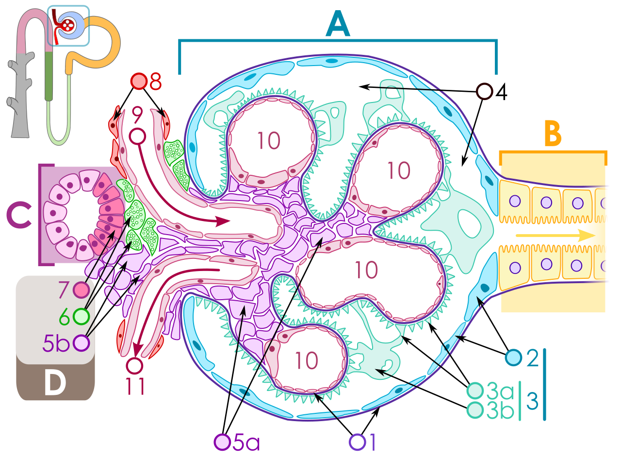
A few landmarks: 8 is the afferent arteriole (blood in) and 11 the efferent arteriole (blood out), the narrower exit that keeps the pressure up. The pale loops (10) are the capillary tuft. A is the filtration wall, with podocytes (1) wrapping the capillaries; B is the start of the tubule. The purple mesh at center (5a/5b) is the mesangium, the tuft’s scaffolding.
Illustration by Michał Komorniczak, via Wikimedia Commons, CC BY-SA 3.0.
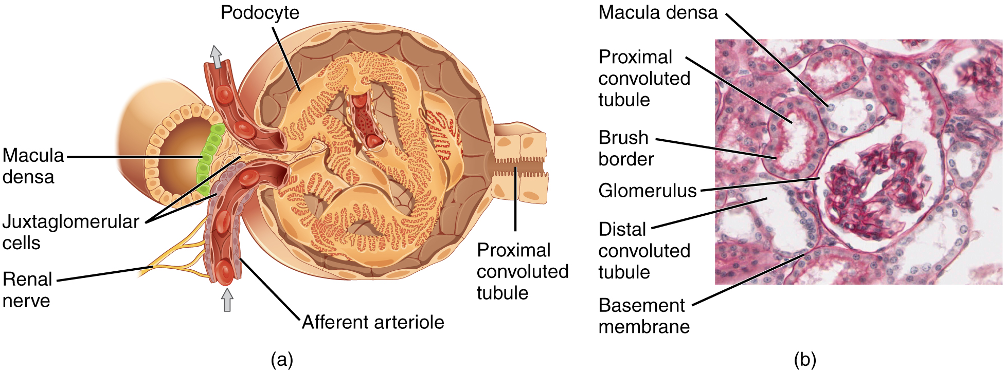
The word-labeled view. Note the afferent arteriole feeding the tuft, the podocytes wrapping it, and the macula densa and juxtaglomerular cells, the pressure-and-salt sensors that release renin. On the right is the same structure on an actual stained slide.
OpenStax College, Anatomy & Physiology, via Wikimedia Commons, CC BY 3.0.
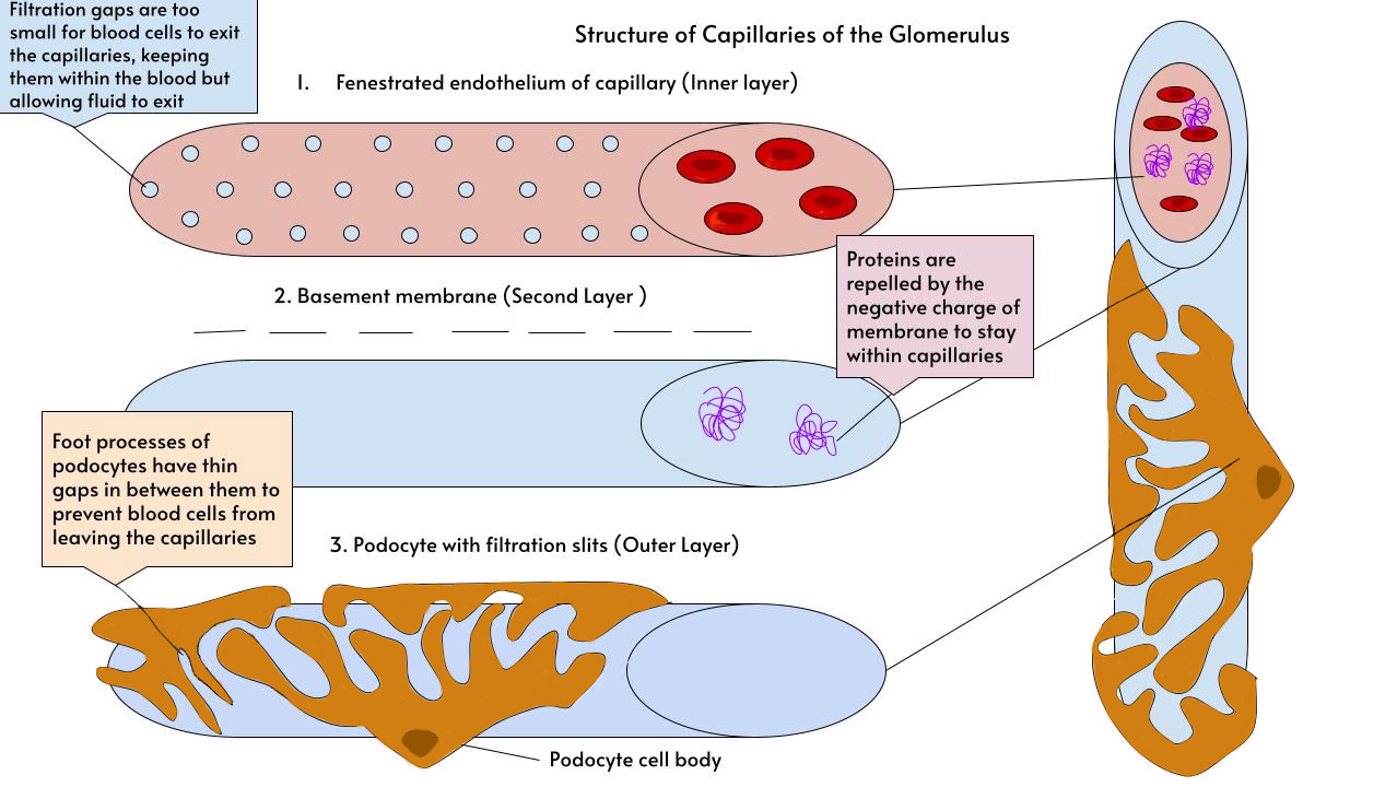
The “only three layers thick” wall from the text: the fenestrated (perforated) inner lining, the basement membrane in the middle, and the podocyte feet with their fine slits on the outside. The notes show the two-part trick, gaps too small for cells, and a negative charge that repels proteins.
Illustration by Alannahlouise, via Wikimedia Commons, CC BY-SA 4.0.
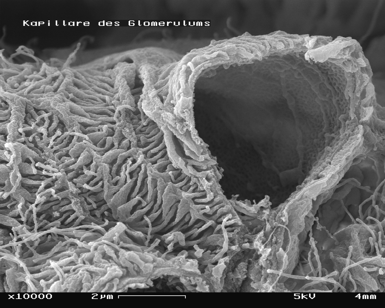
After three diagrams, the actual tissue. A scanning electron microscope image of a mouse glomerulus at 10,000x, one capillary broken open. The ropy, interwoven strands blanketing the surface are the podocyte foot processes, the “interlocking feet” from the text; the perforated surface inside the break is the fenestrated lining. (The German label reads “capillary of the glomerulus.”)
Image by SecretDisc, via Wikimedia Commons, CC BY-SA 3.0.
Ultimately, my job comes down to protecting the millions of these little tangled balls.
Image Credits & Reuse
Fig. 1: Michał Komorniczak, “Renal corpuscle,” via Wikimedia Commons, licensed CC BY-SA 3.0. Original.

Fig. 2: OpenStax College, Anatomy & Physiology, via Wikimedia Commons, licensed CC BY 3.0. Original.

Fig. 3: Alannahlouise, “Structure of the Capillaries of the Glomerulus,” via Wikimedia Commons, licensed CC BY-SA 4.0. Original.

Fig. 4: SecretDisc, mouse glomerulus SEM at 10,000x, via Wikimedia Commons, licensed CC BY-SA 3.0. Original.
